Microscopy and Image Analysis
Microscopy
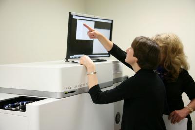
The Molecular Devices ImageXpress 4.0 is used for multicolor fluorescent image-based assays, typically monitoring a protein’s distribution within cells, either from a fluorescent reporter or using immunofluorescent and nuclear staining. The instrument has a robust and highly efficient optical system using a solid state light source and capturing images with a large field of view (5.5 Megapixel) CMOS camera. Laser and software autofocus options enable it to take images from 96- and 384-plates in minutes. It is well suited for a range of assays resulting from five excitation channels and 4X, 10X, 20X, 40X and 60X objectives. In addition, it can take brightfield images. Image deconvolution can be applied during acquisition to sharpen boundaries and define puncta.
Image features can be quantified using predefined image analysis algorithms accessed through Graphical User Interfaces, or through tools enabling a higher level of specification for feature selection. Alternatively, analysis “pipelines” can be developed using CellProfiler, a very powerful and intuitive freeware program.
Image Analysis: Features of complex cellular phenotypes captured in microscopy images can be identified and quantified using high throughput methods. In most cases, nuclei are identified first, and other cellular features dependent upon that nuclei assignment are delineated. Size, shape, signal intensity and number, are just a few of the features which can be analyzed from fluorescent or transmitted light multi-channel images. We use statistical measures on analyses derived from replicate images of control treatments to assess algorithm performance.
The price for developing an algorithm, processing images and analyzing the results is based on a Center use hourly fee, which is subsidized by the Center.
