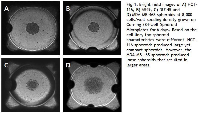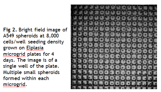
YCMD now has the ability to grow cells in 3D Spheroid culture and screen for cell viability. Laura Abriola (YCMD staff member) and Sydney Wade (summer 2015 undergraduate intern) evaluated different plate types for growing cancer cells in 3D. 3D aggregates of cancer cells—or spheroids—are considered a more accurate representation of the nature of tumors in vivo.
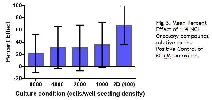
Cell viability was measured using CellTiter-Glo 3D (Promega) or bright field imaging for spheroid size and shape (GE InCell 2200). Spheroid measurements including area, median radius and perimeter were analyzed with CellProfiler image analysis software. Five cancer cell lines known to differ in their ability to form spheroids were seeded at different densities in the different plate types. Plates were evaluated for their ability to form uniform compact spheroids with consistent results from well to well. Two 384-well plates were identified as suitable for screening purposes. Corning Spheroid round bottom plates grew large single spheroids in each well (Fig. 1), and Kuraray Elplasia square microgrid plates grew multiple small spheroids in each well (Fig 2). Next, HCT116 cells grown as spheroids or in 2D were treated with anti-cancer drugs at a single 10 uM concentration and incubated for 3 days, followed by the CellTiter-Glo 3D cell viability assay. Spheroids were grown at different seeding densities to generate different size spheroids.
We found that the multidimensional spheroids were more resistant to traditional anti-cancer drugs in comparison to their 2D counterparts. None of the drugs killed the spheroids more than the 2D grown cells. The mean percent effect for all the drugs together also showed a lower mean value than the 2D grown cells. (Fig 3).